

 GE Healthcare has announced that they have received FDA approval of SenoClaire, GE’s new breast tomosynthesis solution designed with a novel three-dimensional approach to imaging technology. Working with Massachusetts General Hospital, GE developed SenoClaire technology to use a short, low-dose X-ray sweep that moves around a positioned breast and features 9 exposures that are acquired through a unique step-and-shoot approach, which removes any potential motion from the tube that in turn helps to significantly reduce blurry imaging while increasing image sharpness.
GE Seeks to Advance Breast Cancer Screening
Despite the progress that has been made over the past years in advancing mammography technology, GE Healthcare notes that an estimated 20 percent or more of breast cancers are still missed when using digital mammograms. One major reason for the inaccuracies is that cancer can be obscured by normal tissues within the breast. Additionally, normal tissues located in different parts of the breast may become superimposed in the imaging, thus generating a false positive for possible cancer, and leading to wasted time, expense, and stress.
Digital Breast Tomosynthesis (DBT) is a technology that GE is developing to help reduce the inaccuracies and limitations of traditional breast imaging. Developed at Boston’s Massachusetts General Hospital by a leading expert in breast cancer detection and diagnosis, in partnership with GE, SenoClaire is a new DBT solution that received FDA approval last week.
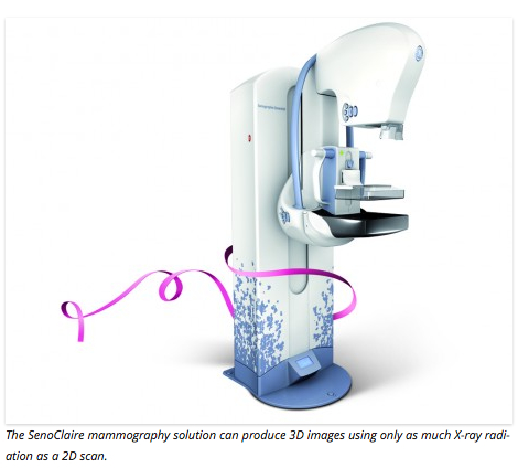
 The SenoClaire mammography solution’s particular distinction is that it is claimed to produce 3D images using only as much X-ray radiation as a 2D scan. GE’s SenoClaire breast tomosynthesis is a three-dimensional imaging technology that uses a low-dose short X-ray sweep around the compressed breast with only nine exposures. It then uses powerful computing to produce multiple images like pages in a book that look like cuts through the breast, so that each plane is virtually free of overlapping tissue in front and behind. This results in sharply rendered views of each plane, while structures in the other planes in front and behind disappear.
SenoClaire, powered by ASiR*DBT, provides superior sensitivity for architectural distortions and masses, giving clinicians more clinical confidence while delivering the same amount of dose as a digital mammography acquisition of the same view.The technology can then reconstruct a representation of the entire breast by combining the set of virtual cuts. Tomosynthesis, which is already in use in imaging solutions such as CT scans and MRIs, is a way to get the three-dimensional information that clinicians need.
A key challenge when performing screening mammography is keeping the radiation levels as low as possible. A single MLO view of SenoClaire, powered by ASiRDBT, provides clinical non-inferiority when compared to 2-view digital mammography, and there is no increase in dose from a 2D standard mammogram to a 3D view, which means there is no increased radiation to patients during a SenoClaire breast exam. In short, it’s half the dose, with one single compression.
[adrotate group=”3″]
 With GE’s SenoClaire, 3D breast screening technology helps clinicians uncover small cancers which can be a limiting factor in standard 2D mammography. “As a radiologist, its important to offer technology like this for patients that produces higher image quality without increasing dose,” says Dr. Murray Rebner MD, FACR, Professor of Diagnostic Radiology and Molecular Imaging at Oakland University William Beaumont School Of Medicine and Director of the Division of Breast Imaging and Intervention at Beaumont Hospital, Royal Oak. “We believe this technology can have a significant impact on helping clinicians to identify breast cancer.”
Due to its low dose without diminished clinical accuracy, SenoClaire has potential to replace digital mammography exams in screening at half the dose to help doctors detect breast cancer. As part of the learning curve process, an additional CC view may be considered along with the 3D MLO view, which will help radiologists become more comfortable with reviewing only the 3D MLO sequence in the near future.
“Our technology provides extremely clear images explains” Remy Klausz, Principal Engineer, Detection and Guidance Solutions, GE Healthcare. “One way of performing DBT would be to have the x-ray tube move continuously taking pictures while in motion. Imagine taking a picture with a camera inside a fast moving car. The pictures would likely turn out blurry. To avoid that blur and capture sharper images, its best to stop the car to take the picture. SenoClaire uses a comparable method called step and shoot, which eliminates this type of blurring. Its different from other 3D tomosynthesis technology, which can result in blurring. Importantly, SenoClaire delivers this exceptional3D image but at the same low-dose radiation level as standard 2D technology.”
 GE researched and developed step-and-shoot digital breast tomosynthesis over the last decade. “This is a key milestone in our mission of providing women with cutting edge screening technology to detect early breast cancer,” says Dr. Daniel Kopans, Senior Radiologist at the Breast Imaging Division Department of Radiology, Massachusetts General Hospital in a release. “When cancer is identified and treated earlier, we know women have a better rate of survival. When cancer is identified and treated earlier, lives can be saved.”
In the step-and-shoot method, after each image is taken, the movement resumes and the x-ray source moves to the next position. The average acquisition takes less than 10 seconds. This approach heightens image clarity and is made possible because of the technology’s lightweight and sophisticated steady rotating tube-arm.
“Tiny calcifications can often be the only indicator of breast cancer. Some devices have to prioritize the speed at which an image is acquired, due to the continuous sweep they employ,” says Dr. Klausz. “And a simple way to increase speed is to reduce the number of pixels, thereby reducing image quality. But GE’s step-and-shoot mode makes this pixel reduction unnecessary, thereby allowing a strong visualization of small objects, such as microcalcifications. In a study from the Massachusetts General Hospital, it was found that microcalcifications were more clearly depicted on GEs DBT system with greater clarity than with 2D digital mammography.”
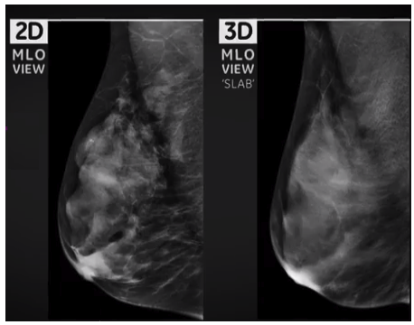
GE Healthcare says SenoClaire enables better characterization of malignant and benign findings by providing a 3D MLO view that has superior detection for architectural distortions and masses compared with 2D FFDM. Clinical results have shown that the recall rate after a 3D MLO examination is lower than the recall rate after a 2D FFDM exam. The SenoClaire detector delivers high DQE at low dose for visualizing microcalcifications without binning, a process that regroups pixels for faster readout speeds and improved signal-to-noise ratios, but with reduced image quality.
 Step-and-shoot tube motion preserves microcalcification sharpness and avoids image blur, since the tube makes a complete stop for each of the nine exposures. This provides higher peak contrast for microcalcifications than traditional continuous tube motion. An anti-scatter solution designed for tomosynthesis, the SenoClaire grid in 3D reduces scattered radiation and improves detectability in 3D for breasts of above-average thickness, while preserving dose and performance.
SenoClaire uses ASiRDBT, an iterative reconstruction algorithm that yields FFDM-like images and positively impacts microcalcification conspicuity versus the traditional Filtered Back Projection (FBP) algorithm.
“Today’s announcement marks a key milestone in our mission of providing women with cutting edge screening technology to detect early breast cancer,” Dr. Kopans observes. “When cancer is identified and treated earlier, we know women have a better rate of survival.”
In addition to offering more clarity, confidence and low dose, SenoClaire offers a complete solution by helping to improve overall workflow. SenoClaire is compatible with Centricity PACS with Universal Viewer and supports the DICOM standard that can be read by capable PACS vendors. When SenoClaire is combined with GE Healthcares Centricity PACS and Centricity Clinical Archive solution, clinicians have access to the patients longitudinal record, providing data that helps to enable better patient care.
 With the FDA’s approval of SenoClaire, we build on our breast care continuum which offers physicians and patients a complete suite of solutions from screening and diagnosis through treatment and monitoring, says Catherine Tabaka, Chief Marketing Officer, GE Healthcare, Detection and Guidance Solutions. “SenoClaire not only offers patients a new solution to help clinicians better detect breast cancer, but does so with low dose radiation and high image quality. This new generation technology, breast tomosynthesis, together with innovative solutions like contrast enhanced spectral mammography, automated whole breast ultrasound, and molecular breast imaging will equip healthcare providers with a comprehensive set of tools that will help their patients across the entire breast care continuum.”
The Breast Imaging program at Massachusetts General Hospital Imaging provides comprehensive screening and diagnostic exams using the latest technology, coupled with the expertise of one of the country’s largest teams of specialized breast radiologists.
Working through the Comprehensive Breast Center located at Mass. General in Boston, as well as at several community locations, the Breast Imaging team focuses exclusively on the detection and diagnosis of breast cancer using state-of-the-art mammography including 3D mammography (breast tomosynthesis), ultrasound, and MRI technology.
The Mass. General Breast Imaging Program includes a team of 12 dedicated breast radiologists from the Breast Imaging division of the Mass. General Department of Radiology, experts in detecting breast lesions through the use of digital mammography, ultrasound and MRI. Working from the Avon Foundation Comprehensive Breast Evaluation Center on the Mass. General Main campus, this group is one of the nation’s largest breast-imaging teams.
A life-long advocate of mammographic screening to increase early detection of breast cancer, Mass. General’s Dr. Daniel Kopans was one of the few experts to challenge the National Cancer Institute’s controversial 1993 assessment that women under 50 did not benefit from regular mammograms. Arguing that the assessment was based on inconsistent studies, he insisted that women in their 40s can significantly benefit from mammograms.
Dr. Kopans’ leadership played a major role in reversing NCI’s assessment, thereby increasing younger women’s access to screening. Since 1990, the breast cancer death rate in the United States has decreased by almost 30 percent, primarily because of an increase in mammography screening.
Dr. Kopans has pioneered innovations in early breast cancer detection, including tomosynthesis, a procedure that increases the accuracy of mammograms without significantly increasing radiation exposure, and the Kopans Wire, a tool that helps clinicians detect small breast tumors.
Sources:
GE Healthcare
Massachusetts General Hospital
Oakland University William Beaumont School Of Medicine
Image Credits:
GE Healthcare