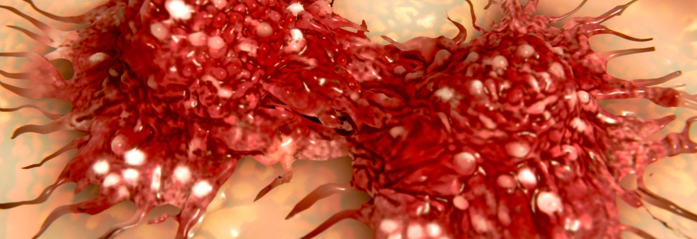
When tumor cells become too densely packed, they start developing blood vessel-like structures — an early step in the process of cancer metastasis, researchers at the University of California, San Diego (UCSD) discovered in a recent study.
The work adds a new level of understanding in the processes that guard cancer spread which may, in the future, allow physicians to assess the risk that a tumor will spread — tailoring the treatment to better contain that risk.
It also paves the way for the development of drugs that target the newly discovered machinery to stop tumors from spreading.
“We are good at targeting tumor growth, but we do not know enough about metastasis,” Stephanie Fraley, a professor of bioengineering at UCSD and the study’s senior study, said in a press release.
Researchers have observed the vessel-like structures for some time, the research team explained in their report, published in the journal Nature Communications. But they did not know which factors triggered their development.
The phenomenon, called vascular mimicry, is linked to some of the most aggressive cancers known.
Researchers of the study, “3D collagen architecture induces a conserved migratory and transcriptional response linked to vasculogenic mimicry,” set out to explore potential factors in the tumor microenvironment, and found that tumor density is of key importance.
When the environment in the tumor became too confined, the cells turned on a specific set of genes, the team discovered. The activation of the genes was linked to patient survival and cancer metastasis in people with breast and a range of other cancers.
The discovery makes it possible for physicians to assess if these genes are active, and therefore gain insight into whether the tumor is aggressive or not. This, in turn, may impact what treatments they choose for a particular patient.
The team came to the conclusion that a constrained environment triggers the formation of the blood vessel-like structures after studying lab-grown cancerous cells in different types of three-dimensional matrix structures.
They believed — based on previous knowledge of how such cells behave in normal lab dishes — that their experiments would show that a denser matrix would block the cells’ spread. But they were in for a surprise.
“We thought that putting cells into this more constrained environment would prevent their spread,” said Daniel Ortiz Velez, the study’s first author and a PhD student in Fraley’s lab. “But the opposite happened.”
A flat lab dish poorly reflects a tumor’s natural environment, a factor that turned out to be quite important.
“It’s critical to have the cells surrounded by a 3D environment that mimics what happens in the human body,” Fraley said.
When cancer cells grew in a more constrained matrix, a specific set of genes were turned on. “It’s almost like the matrix is encoding the gene module,” she added.
To confirm that these genes were really involved in the spread of cancer, the team looked for their presence in databases of cancer gene activity in human tissue samples. It turned out that the presence of these genes was linked to a strong likelihood that the cancer would metastasize aggressively — also when they took other factors, such as a patient’s age, into account.
The team is now validating their findings in cancer animal models and additional databases of human tumors. They also plan to look for ways to prevent the cells from switching into metastasis mode.

