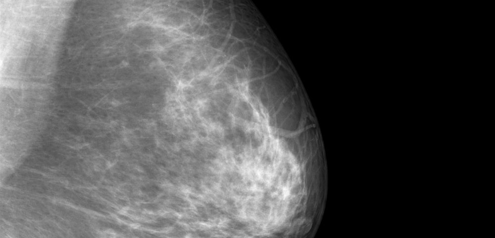
Researchers at California Institute of Technology (Caltech) developed a new imaging technology for breast cancer screening that can scan the whole breast and find tumors within 15 seconds.
The method, called single-breath-hold photoacoustic computed tomography (SBH-PACT), shines a laser light on the breast and detects changes in ultrasonic vibrations. It may one day replace mammograms, according to researchers.
Their study reporting these findings, “Single-breath-hold photoacoustic computed tomography of the breast,” was published in Nature Communications.
Screening for breast cancer is important for an early diagnosis, which has been shown to increase overall survival of patients. But many women avoid mammograms in their routine checkups because the tests are uncomfortable.
While mammograms are a useful imaging technique for early cancer detection, they also expose patients to X-ray radiation. Also, breasts are painfully squished between two plates to flatten the tissue, so that X-rays can penetrate more easily and produce a better image.
Besides the discomfort involved, mammograms have other significant downsides, such as the difficulty in generating clear images in women with dense breast tissue and the tendency to generate false-positive diagnoses.
Researchers at Caltech believe they developed a better alternative: a new imaging method based on a laser-sonic scanner that shines pulses of light into the breast and is able to find tumors within 15 seconds.
Their SBH-PACT method works by shining near-infrared light pulses into the breast, which are then absorbed by hemoglobin molecules inside the patient’s red blood cells.
When these molecules vibrate, vibrations travel through the tissue and are picked up by ultrasonic sensors placed on the patient’s skin. The sensors then convert these data into a detailed image of the breast’s internal structures, which could be as small as a quarter of a millimeter in size and at a depth of four centimeters.
Mammograms aren’t able to provide the contrast in soft-tissue with the level of detail observed from PACT imaging, Lihong Wang, Caltech’s Bren Professor of Medical Engineering and Electrical Engineering, said in a Caltech news story written by Emily Velasco.
In a PACT scan, a woman lies face down on a table while placing one breast at a time on a small recess with the laser scanner underneath. Scanning the entire breast takes approximately 15 seconds, meaning that a clear image can be obtained while women hold their breath.
“This is the only single-breath-hold technology that gives us high-contrast, high-resolution, 3-D images of the entire breast,” Wang said.
Besides high resolution, scan speed is also better with PACT over other imaging techniques, such as magnetic resonance imaging (MRI), which can take up to 45 minutes.
Another advantage of PACT is that unlike MRI, it does not require patients to be injected with contrast agents that can have adverse effects.
“Gadolinium, for example, is a common contrast agent for MRI which is not totally innocuous,” Wang said. “Gadolinium may cause nausea and vomiting, and can remain in the brain for years, with unknown long-term effects. In comparison, PACT is entirely safe; the blood serves as an intrinsic contrast agent, and the laser exposure is well within the safety limits.”
In a recent pilot study, Wang’s team scanned breast cancer patients with different breast sizes and skin pigmentation using PACT, and correctly identified eight out of the nine tumors present without having to resort to ionizing radiation or exogenous contrast.
Now that PACT is already licensed, Wang plans to commercialize it and conduct additional large-scale clinical trials to test its effectiveness as a screening and early diagnostic tool.
“Our goal is to build a dream machine for breast screening, diagnosis, monitoring, and prognosis without any harm to the patient,” he said. “We want it to be fast, painless, safe, and inexpensive.”

