
 Researchers at the University of California, Los Angeles (UCLA) have harnessed the power of large and expensive microscopes for cancer detection into the world’s first lens-free microscope. The device can detect cancerous alterations in a single cell with a high degree of accuracy, as tested in cancerous breast, cervical, and blood cells.
“This is the first time tissue samples have been imaged in 3D using a lens-free on-chip microscope,” said Dr. Aydogan Ozcan, whose lab developed the device, in a news release. “This is a milestone in the work we’ve been doing.”
The technology behind this power is a silicon chip similar to those found in smartphones and digital cameras. Yet the images this device captures do more than snap memories on-the-go: they are capable of identifying abnormalities at the cellular level. “Our microscope provides the same level of quality as a state-of-the-art optical light microscope, and it has a significantly larger field of view, a simpler design, and it is more cost-effective,” added Dr. Ozcan in a UCLA news release on Los Angeles Times.
As senior author of “Wide-field Computational Imaging of Pathology Slides Using Lens-free On-chip Microscopy,” published in Science Translational Medicine, Dr. Ozcan described the technology in detail. In addition to the silicon chip, a light source and sample holder are required. “The bread and butter of this project is a CCD or CMOS imager, which is the same thin chip you find in every digital camera, whether it’s a high-end SLR or a cellphone camera,” explained Dr. Ozcan.
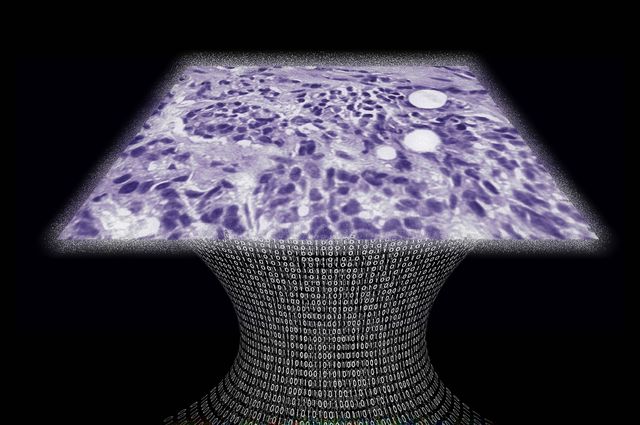
When light shines on the slide holding the sample, the slide projects a shadow onto the imaging chip. As the sample comes closer to the imaging chip and further from the light source, the shadows of cells within the sample become more defined.
Looking at the projected shadow bears no resemblance to looking at the sample through a light microscope. Rather, the image collected by the chip is a murky-looking holograph, which is then reconstructed with software developed in Dr. Ozcan’s laboratory. “The hardest part was creating the computational transformation that takes those nasty-looking shadowy patterns and give you the truth of what is happening,” said Dr. Ozcan. “That was the computational puzzle.”
[adrotate group=”3″]
During testing, breast cancer was detected with an overall accuracy of almost 99% when compared to detection by a pathologist’s clinical evaluation with a light microscope. The goal is to someday equip resource-limited clinicians with point-of-care diagnostics such as these that can make life-saving diagnoses. “A small nurse’s office that doesn’t have a pathologist on staff could transmit digital images created by our microscope to an expert in another city, or another country,” described Dr. Ozcan. “Mobile health and global health is where I would like to channel the things we create.”
Before this diagnostic is put into practice, the team has more work to do. “For other people to use it, it needs to be like Windows,” stated Dr. Ozcan, indicating that the computer software that allows digital image reconstruction needs to be made more user friendly. “You can think of our interface as a very early version of the personal computer, where you have to write code to do anything.” Fortunately, the team is excited by the technology and will continue to develop their lens-free microscope to detect breast and other cancers.