
 One of Scottsdale-based Arizona Breast Cancer Specialists’ researchers, Robert Kuske, MD, presented data at the annual meeting of the American Society for Therapeutic Radiology and Oncology (ASTRO) held September 14-17 that suggests a new surgical marker can increase the preciseness of delivered radiation therapy by radiation oncologists to breast cancer patients. The presentation focused on the BioZorb™ Tissue Marker from Focal Therapeutics, Inc.
“Like many great innovations, the design of the new marker is relatively simple and its effects are profound,” said Dr. Kuske, MD, in a press release provided by the company. “The reduction in treatment volumes means that with this marker, women have far less risk of damage to healthy tissue during treatment, and that they can expect better cosmetic results.”
These findings were made through two main studies. One study documented 51 cases of physician’s clinical experience with BioZorb. Using the surgical marker enabled smaller volumes of radiation treatment and doses in patients receiving either boost or accelerated partial breast irradiation (APBI), a technique pioneered by Dr. Kuske to reduce the time of treatment from six weeks to five days for patients with select types of early-stage breast cancer.
Another study used BioZorb alongside oncoplastic surgery. “The marker proved to have a unique ability to complement oncoplastic techniques,” said Linda Ann Smith, MD, a breast surgeon at Comprehensive Breast Care who authored the study. “This is due to its three-dimensional, open spiral shape. Our research showed that the marker improves cosmetic results across the board. This is especially true when combined with reconstructive oncoplastic surgery.”
One of the challenges of oncoplastic surgery is the need to rearrange patients’ tissues when trying to maintain breast shape following surgery. By placing the marker in the breast, radiation site visualization is enhanced over the level attainable by using traditional markers.
[adrotate group=”3″]
For one patient in particular, Charmazel Dudt, treatment was greatly enhanced. “Dr. Smith told me that normally, she would have been worried about my receiving radiation because my cancer was located near my heart and I have two stents,” said Dudt. “But using this new marker made her much less concerned because my treatment was going to be very precise. That meant there was less risk of radiation damage to my heart. And, of course, that’s just how things turned out. I’m so grateful.” Dudt first had the tissue marker placed by Dr. Smith during a lumpectomy. Then, she left for treatment by Dr. Kuske for an interstitial brachytherapy procedure, which is a multi-catheter APBI technique shown to reduce the average number of catheters required during surgery.
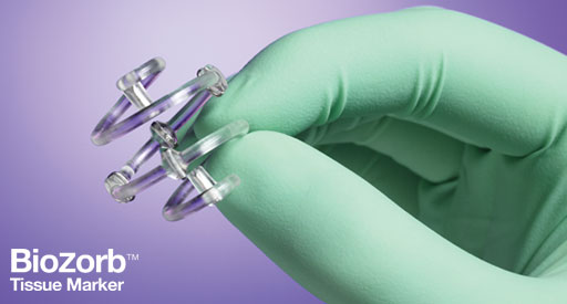
In addition to Dudt, a number of other women in the United States and New Zealand have benefited from the unique, open-spiral surgical marker. The marker consists of six titanium clips permanently fixed in a 3D array to provide a landmark at the excised tumor site. However, after the marker has served its purpose as a securing point during oncoplastic reconstruction, positioning, and planning, it can be bioabsorbed by the body slowly over time, preventing the need for a follow-up surgery and further enhancing treatment of breast cancer patients.