 A new study entitled “Engineering Gold Nanotubes with Controlled Length and Near-Infrared Absorption for Theranostic Applications” details the successful use of gold nanotubes in both in vitro an in vivo settings as a potential therapeutics to human diseases, such as cancer. The study was published in the journal Advanced Functional Materials.
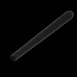
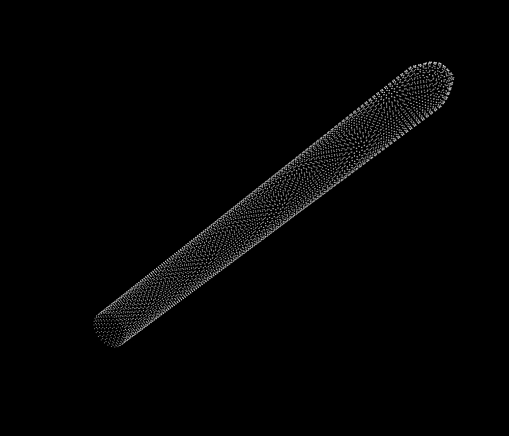
In this new study, a team of researchers at the School of Physics and Astronomy, the Leeds Institute for Biomedical and Clinical Sciences and School of Electronic & Electrical Engineering, University of Leeds developed gold nanotubes (NTs), i.e., gold tubular nanostructures, with a series of new characteristics to enhance their diagnostics and potential therapeutics applications. First, the authors developed a technique to control the length of the tubes and created nanotubes with a well-defined shape. These dimensions then allowed the tubes’ absorption of a specific type of light, in the ‘near infrared’ region. The development of these features is essential for therapeutics, such as Photothermal therapy — this is a minimally invasive procedure where photothermal conversion agents (PTCAs) have the power to absorb light and convert it into heat — and is used to specifically ablate cancer cells. Underlying this and other similar approaches is the absolute necessity of tubular nanostructures to absorb in the near-infrared and have low toxicity. For the latter point, the team coated the nanotubes with poly (sodium 4-styrenesulfonate) (PSS) to ensure its low toxicity.
The team tested the new developed gold nanotubes in mice, where they injected the nanostructures intravenously. The imaging of the nanotubes inside the body was allowed by using a new imaging technique, known as ‘multispectral optoacoustic tomography’ (MSOT). The authors had previously tested the structures in vitro with cells and observed that colorectal cancer cells readily uptake the nanotubes effectively inducing cancer cells photo thermal ablation.
In vivo, the authors showed that nanotubes are excreted from the body, establishing their potential as effective and safe therapeutics to use in a future clinical setting. The study is the first showing the potential of nanotubes, both in vitro and in vivo.
Dr Sunjie Ye, School of Physics and Astronomy and the Leeds Institute for Biomedical and Clinical Sciences and study first author commented in a press release, “When the gold nanotubes travel through the body, if light of the right frequency is shone on them they absorb the light. This light energy is converted to heat, rather like the warmth generated by the Sun on skin. Using a pulsed laser beam, we were able to rapidly raise the temperature in the vicinity of the nanotubes so that it was high enough to destroy cancer cells.”
Dr James McLaughlan, at the School of Electronic & Electrical Engineering, University of Leeds added, “This is the first demonstration of the production, and use for imaging and cancer therapy, of gold nanotubes that strongly absorb light within the ‘optical window’ of biological tissue. The nanotubes can be tumor-targeted and have a central ‘hollow’ core that can be loaded with a therapeutic payload. This combination of targeting and localized release of a therapeutic agent could, in this age of personalized medicine, be used to identify and treat cancer with minimal toxicity to patients.”

